The examination of scar cancer primarily involves a series of medical examinations to determine the nature and extent of the lesion. Scar cancer, also known as keloid skin cancer or scar tissue cancer, is a rare type of skin cancer that typically occurs in areas where scar tissue has been present for a long time. The methods for examining scar cancer mainly include clinical examination, imaging examination, and pathological examination.
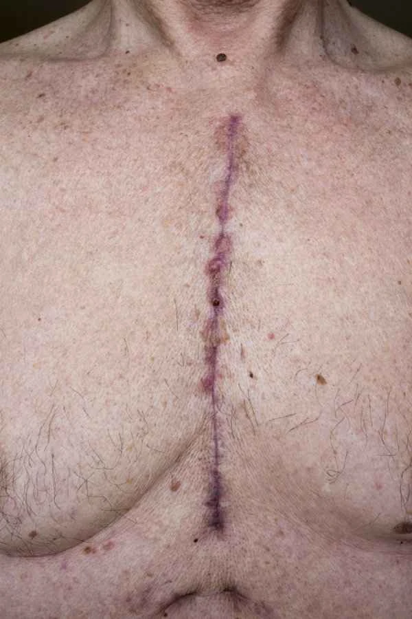
During a clinical examination, doctors assess the appearance and texture of the lesion area through visual inspection and palpation, noting its color, size, shape, and the presence of ulcers. Imaging studies, such as ultrasound, CT, or MRI, help doctors understand the depth and extent of the lesion, as well as whether lymph node metastasis is present. Finally, pathological examination is the gold standard for diagnosing scar cancer. This typically involves obtaining a tissue sample through a biopsy, which is then examined under a microscope by a pathologist to determine the nature of the lesion.

In addition to the examination methods mentioned above, it is important to note that the diagnosis of scar cancer requires a comprehensive consideration of medical history, clinical manifestations, and examination results. During the examination, doctors will utilize multiple diagnostic tools to ensure accuracy. Because scar cancer is relatively rare, the diagnostic process may present some challenges, such as differentiating it from other skin lesions. Patients are advised to seek medical attention promptly if they experience symptoms suggestive of scar cancer and to undergo examination and treatment under the guidance of a professional physician.

[Management Tip:]
1. Have regular skin checkups, especially for areas with long-term scarring.
2. Pay attention to changes in the skin, such as abnormalities in color, size, or shape.
3. If any suspicious symptoms appear, seek medical attention promptly for a professional examination.